
Cardiological Risk Assessment
-
A Online Grayscale Feature Classification Paradigm for Cardiological Risk Assessment on Diabetic Cohort using IVUS
Tadashi Araki, Nobutaka Ikeda, Nilanjan Dey, Sourav Samanta, Ayman El-Baz, Filippo Molinari, Elisa Cuadrado Godia, Luca Saba, Andrew Nicolaides, Jasjit S Suri.
Physics in Medicine & Biology. 2014. (Under Review) [Impact Factor: 2.701]
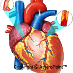
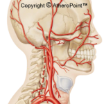 Abstract: The study of automatic determination of cardiological risk assessment is based on Support Vector Machine (SVM) classification approach which uses shape-based coronary calcium lesion features and neurological risk biomarker cIMT. The study was undertaken on a cohort of 100 patients. Acquisition of IVUS data was done by using a 40 MHz Atlantis SR Pro, Boston Scientific intravascular ultrasound catheter. A reverse engineering strategy was adopted for automatic detection and segmentation of coronary calcium lesions in IVUS video frames. Different grayscale based features of the detected lesion such as (i) Haralic texture features, (ii) Gabor features, (iii) fractal features (Total 17 features), were computed for risk classification. A novel, automated and 510 (K) FDA cleared software; AtheroEdge™ (AtheroPoint LLC, Roseville, CA, USA) was used for computation of cIMT on B-mode carotid ultrasound. Our results demonstrate that the shape-based coronary calcium lesion characteristic features are capable of accurately classifying 85% of neurological risky patients. This analysis reports classification of neurological risk biomarker through carotid window (carotid imaging) called intima-media thickness (IMT).
-
Cardiological Risk Assessment using Support Vector Machine by combining Coronary Calcium Lesions and cIMT as a Neurological Risk Biomarker
Luca Saba, Tadashi Araki, Nobutaka Ikeda, Nilanjan Dey, Suvojit Acharjee, Ayman El-Baz, Filippo Molinari, Elisa Cuadrado Godia, Andrew Nicolaides, Jasjit S Suri.
Ultrasound in Medicine and Biology. 2014. (Under Review) [Impact Factor: 2.455]
Abstract: The study of automatic determination of cardiological risk assessment based on SVM classification approach using shape-based coronary calcium lesion features and neurological risk biomarker cIMT. A study was undertaken on a cohort of 100 patients. Acquisition of IVUS data was done by using a 40 MHz Atlantis SR Pro, Boston Scientific intravascular ultrasound catheter. A reverse engineering strategy was adopted for automatic detection and segmentation of coronary calcium lesions in IVUS video frames. Different shape-based characteristic features such as (i) mean lesion thickness, (ii) mean standard deviation of lesion thickness, (iii) mean central line length, (iv) mean length of all branches, (v) mean span (arc angle) of the detected lesion, (vi) mean lesion irregularity and (vii) lesion distance to catheter center were computed for risk classification. A novel, automated and 510 (K) FDA cleared software; AtheroEdge™ (AtheroPoint LLC, Roseville, CA, USA) was used for computation of cIMT on B-mode carotid ultrasound. Our results demonstrate that the shape-based coronary calcium lesion characterization and classification software is capable of accurately classifying 80% of cardiological risky patients (sensitivity is 0%, specificity is 80%). This analysis reports classification of cardiological risk patients by combining coronary calcium lesions from IVUS and neurological risk biomarker of carotid IMT is able to classify coronary artery disease patients with high success rate. -
Hunting for Calcium Lesions in Intravascular Ultrasound: an automated and accurate calcium volume quantification and its correlation to automated cIMT using AtheroEdge
Tadashi Araki, Nobutaka Ikeda, Nilanjan Dey, Suvojit Acharjee, Filippo Molinari, Luca Saba, Ayman El-Baz, Andrew Nicolaides, Jasjit S Suri.
Medical Physics. 2014. (Under Review) [Impact Factor: 2.83]
Abstract: The carotid intima-media thickness (cIMTs) using AtheroEdge™ is a strong predictor of cerebrovascular complications. Our recent studies showed a strong correlation of cIMT with Ankle Brackle Index (ABI) and Syntax Score. In our previous research work we have reported that the normalized volume of the detected coronary calcium from IVUS videos using shape-based approach correlates to cIMT. We here demonstrate an improved method of false positive calcium elimination thereby improving the automated calcium volume estimation followed by the correlation with cIMTs. An automated computer-based application is developed and tested on 92 IVUS patient volume videos. The concept is to estimate the calcium regional area by counting all the pixels of the region corresponding to the coronary calcium in each detected frame. To segment the calcium region, a reverse engineering strategy is adapted, that is removal of non-region of interest which consists of media/adventitia high gradient regions along with catheter regions. The calcium is automatically detected once these non-regional interest regions are removed. False positive calcium elimination is corrected using a combination of textured entropy and distance constraint. Volume is then quantified by summing all the pixels in the corrected segmented lesion in automatically detected video frames. Normalized calcium volume is then compared to AtheroEdge™-cIMTs automatically taken from the B-mode carotid ultrasound using Pearson correlation coefficient.The software is able to detect the calcium lesion frames accurately and automatically, and also computes the corresponding calcium volume. Further, it establishes a relationship between calcium volume and cIMTs estimated by AtheroEdge™ from AtheroPoint™. We show a 6.16% improvement in elimination of false positive using our textured entropy strategy. Further, our results show a mean improvement in correlation coefficient between calcium volume and AtheroEdge™-cIMT by 45.42% when taking both the left and right carotids using our new method of textured entropy and distance constraint. Textured entropy strategy improves the correlation between coronary calcium volume computed from IVUS using shape-based approach and cIMTs computed using AtheroEdge™ software from carotid ultrasound. The system is fully automated and links cardio and neuro-fields having same genesis as atherosclerosis genesis.